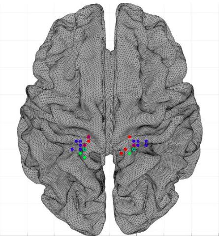
Precise location of the area of the sensory cortex devolved to the clitoris
En ayant recours à l’imagerie par résonance magnétique fonctionnelle (IRMf), des chercheurs allemands ont localisé avec précision la zone du cortex cérébral dévolue à la sensibilité génitale de la femme. Pour ce faire, ils ont analysé, chez vingt femmes, la réponse neuronale à une stimulation expérimentale du clitoris. Leurs résultats ont été publiés en ligne le 10 décembre 2021 dans The Journal of Neuroscience.
We know that the sensitivity of the whole body projects itself at the level of the parietal lobe of the cerebral cortex.In 1950, Wilder Penfield and Theodore Rasmussen, researchers from McGill University in Montreal, established a detailed cartography of the cerebral cortex thanks to electrical stimulation made point by point during neurosurgical interventions.They thus drawn what we call the sensitive or sensory homonculus.
Represented with an oversized hand and face, the sensitive homonculus corresponds to a silhouette representative of the way in which the different regions of the human body project at the level of the cerebral cortex.The zone of the cortex devolved to sensitivity, which receives in particular the main sensory influxes of the surface of the body, is called the primary somatosensory cortex (primary sodetheical area S1).This is located in the ascending parietal circumvolution (post-central gyrus) and includes regions called Brodmann 1, 2, 3 (3A and 3B) areas.
In 2005, thanks to a technique based on stimulation of the penis by a toothbrush with very flexible hair, a German team located the sensitive cortical representation of the human penis between the legs and the trunk of the sensory homonculus.It therefore remained to drive in women an experience of stimulating the clitoris to locate female genital representation at the level of the sensory cortex.
Stimulation of the clitoris via a vibrant membrane
In order to precisely locate the sensory cortical representation of the clitoris, the researchers used functional MRI.This made it possible to visualize neural activity during clitoral stimulation.
The participants had to wear a stimulation device fixed by a velcro flexible belt on underwear.The clitoris region was stimulated in a non -invasive manner by a small membrane oscillating under the effect of an air flow.This membrane was fixed under Mount de Venus in the clitoris region.
Genital tactile stimulation alternated with stimulation of the right hand, as a comparison.Four series of stimulation was carried out, including eight clitoris stimulations and eight others on the hand, each lasting 10 seconds alternating with a rest of 10 seconds.Twenty women participated in this study conducted by Christine Heim from the Charitable University Hospital (Berlin).After each series of stimulation, the participants had to indicate the intensity of the pleasant feeling or sexual excitement on a visual scale.
In this study, the stimuli of the clitoris did not consist, as in other protocols used previously, in the electrical stimulation of the dorsal nerve of the clitoris clitoris.The experience did not imply the touch of areas located near the clitoris.Finally, tactile stimulation has not caused significant sexual excitement as when stimulation is applied by the person himself or a partner.We can note in this regard that the touch stimulation issued by the experimental device placed on the underwear has never been felt as unpleasant, no more than it caused intense sexual excitation.

Functional MRI has made it possible to delimit the most activated cortex (vertices) points within the right and left hemisphere when stimulating the clitoris and the back of the hand of the hand stimulation.The researchers also determined the average thickness of the cortical surface containing the 10 functionally the most active points in each hemisphere.
Participants, an average age of 23 years, were also questioned about the frequency of their intercourse in the last twelve months as well as since the start of their sexual life.The majority of these women were heterosexual (17) or bisexual (3).They were all at different times in their menstrual cycle during the study.The average frequency of intercourse in the past year was 1.9 per week.
The results of the functional MRI show that tactile stimulation of the clitoris region has induced a significant neuronal activation, located in the S1 sometheical area of the two hemispheres.More specifically, in all women, neural activation occurred in the areas of Brodmann 1, 2, 3 (3a and 3b)*.Neural activation peaks in response to clitoris stimulation varied considerably between women.It turns out, however, that individual location within the sensory s1 cortex varies, with activation peaks that vary clearly between each woman.
Correlation between the thickness of the activated cortical area and sexual activity
Researchers indicate that the thickness of the activated cortical area is greater in women with more sexual intercourse.There is thus a significant correlation between the thickness of the cortex at the level of the region activated in the left hemisphere and the frequency of sexual intercourse in the last twelve months **.
Likewise, a greater frequency of relations since the beginning of sex life was significantly correlated with the thickness in the left hemisphere of the cortical area of representation of the genitals.This lateralization on the left side is very astonishing insofar as the cortical sensitive representation of the clitoris is bilateral.Researchers cannot provide explanations but point out that the decrease in thickness of the sensory cortex after sexual abuse is limited to the left hemisphere.
Post blog: How to recognize hpv in men (human papillomavirus) - http: // t.CO/4VFHFJIWBC
— gaylifeafter40 Wed Dec 17 23:04:31 +0000 2014
Nothing such has been observed concerning the representation of the hand at the cortical level.In other words, the thickness of the area devoted to the sensitivity of the hand was not significantly associated with the frequency of sexual intercourse in recent months or since the beginning of sex life.This therefore confirms the existence of a specific association between the touch of the clitoris and the thickness of the cortical zone devolved to genital sensitivity.
Sensory brain plasticity
This study is the first to report that the thickness of the cortical area representing the genital region of women in the somato-sensory cortex varies according to the frequency of sexual intercourse during the past year and since the start ofSex life.This suggests the existence of a neural plasticity which depends on sexual practice, according to the general principle according to which plasticity is an adaptive response, conditioned for the use of the function, which the Anglo-Saxons summarize by speaking of use-it-or-lose-it (used or lost).
This cerebral plasticity depends on the nerve impulses that reach it.And German researchers to point out that they have previously observed that the thickness of the sensory cortex devolved to the genitals is reduced in adult women who have suffered sexual abuse during childhood.This result, published in 2013 in the American Journal of Psychiatry, could indicate that highly aversive and inappropriate stimulation can influence the sensory cortical representation.Brain plasticity could then play a protective role for the child victim of sexual abuse.
According to the authors, several mechanisms could contribute to the brain plasticity associated with the sensitive cortical representation of the genitals: formation of new synapses, growth of dendrites, increase in myelinization, reinforced connectivity between thalamus and somatosensory cortex.
Female genital representation in the homonculus
These tactile stimulation experiments indicate that the representation of the clitoris at the level of the sensory cortex is located near that of the hips and the upper part of the legs.
The first representation of the Penfield and Rasmussen sensory homonculus of 1950 indicated that the area devoted to male genital sensitivity is below that of the foot.
The silhouette of the female homonculus therefore differs from that of the male homonculus.Now it remains to draw it.
Marc Gozlan (follow me on Twitter, Facebook, Linkedin)
* Bilateral localized activation located in S1 within the dorso-lateral part of the postcentral gyrus.
** No significant correlation has been observed in the right hemisphere, which suggests the existence of a structural variation only on one side of the brain.There was no significant difference in thickness of this area according to the moment of the menstrual cycle.
To find out more: Knop AJJ, Spengler S, Bogler C, and Al.Sensory-tactile Functional Mapping and Use-associated Structural Variation of the Human Female Genital Representation Field.J neurosci.2021 Dec 10: JN-RM-1081-21.Doi: 10.1523/Jneurosci.1081-21.2021
Michels L, Mehnert U, Boy S, et al.The somatosensory representation of the human clitoris: an fmri study.Neuroimage.2010 Jan 1; 49 (1): 177-84.Doi: 10.1016/J.neuroimage.2009.07.024
Kell ca, von kriegstein k, rösler a, et al.The sensory cortical representation of the human penis: revisiting somatotopy in the male homunculus.J neurosci.2005 Jun 22; 25 (25): 5984-7.Doi: 10.1523/Jneurosci.0712-05.2005
Heim cm, mayberg hs, mletzko t, nemeroff cb, pruessner jc.DECREASED CORTICAL REPRESENTATION OF GENITAL SOMATOSOSORY FIELD After Childhood Sexual Abuse.Am J Psychiatry.2013 Jun; 170 (6): 616-23.Doi: 10.1176/APPI.AJP.2013.12070950


